Prions: A Lurking Threat
Introduction
A recent discovery at a South Carolina beef processing plant raised new questions about a poorly understood known for causing swift onset dementia and the buildup of amyloid fibers in the brain that cause Swiss cheese like damage to humans. So, what is this mysterious pathogenic agent? Prions are proteinaceous infectious particles that were first identified approximately 66 years ago and were not officially named or isolated until 1982. Prion diseases are transmissible spongiform encephalopathies that cause the malfunction and abnormal folding of prion proteins (PrP). As a result of this misfolding, proteins begin to clump together in the central nervous system, primarily the brain, leading to irreversible damage to neurons. Currently, all prion diseases remain incurable and fatal, usually within one year of diagnosis. Prion disease may also be a result of inherited polymorphisms in the PRNP gene that encodes for the prion protein (PrP), spontaneously, or through contaminated food sources such as beef. Both animals and humans are susceptible to these diseases and the most common variations are described in Table 1. In this blog, we will describe prion diseases, how they impact both human and animal health, and the challenges that need to be overcome to successfully decontaminate these hardy pathogenic agents.
Impact of Prions on Human and Animal Health
Infectious prions are unique pathogenic agents as they are solely composed of proteins and lack nucleic acids. When disease occurs, traditional prion proteins (PrP) reform into an abnormal sequence (PrPSc).1 They have the distinctive ability to recruit undiseased proteins in the brain, causing them to misfold, often acting as a template for others.1 This recruitment allows for rapid and progressive neurological damage as the proteins begin to clump together. While prion disease can be found in both humans and animals, they often emerge differently. For example, humans are most impacted by progressive dementia, while animals often present with ataxia.1 Prion diseases are associated with a long incubation period compared to most pathogenic agents ranging from about 15 months to over 50 years depending on the disease. Approximately 300 cases are diagnosed every year in the United States alone. One study discusses the link between incubation period and brain weight.2 Furthermore, the study suggests that animals with lower brain weights may experience more virulent strains of disease.2 Larger brains are believed to result in a longer incubation period because it “reduces the concentration of pathogenic prions by more solvable capacity leading to the decrease of attachment rate of prions to existing aggregates”.2
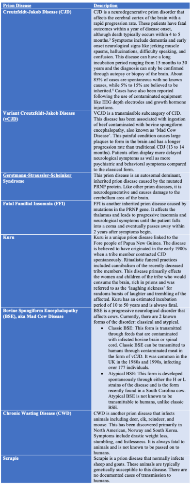
Creutzfeldt-Jakob Disease (CJD) remains one of the most common neurodegenerative prion diseases in the world. The physical manifestations and etiology of the disease are described in depth in Table 1. While most cases occur spontaneously, iatrogenic (caused by medical exposure) forms of the disease have been described in the literature, originating from contaminated medical supplies. Before widespread knowledge of prions’ place on the pathogen hierarchy, contaminated surgical and diagnostic equipment like EEG depth electrodes and dura mater grafts led to outbreaks of prion diseases in otherwise healthy individuals.1 Growth hormone injections in the 1980s became contaminated after harvesting from deceased individuals infected with a prion disease, leading to the deaths of around 85 individuals.3 This helped prompt the shift to synthetic growth hormone and more efficacious sterilization and disinfection methods.
In line with current events, a cow from Tennessee that had been transported to a beef processing plant in South Carolina recently tested positive for bovine spongiform encephalopathy (BSE or Mad Cow Disease). Upon arrival, it began to demonstrate symptoms of the disease and was immediately segregated and euthanized before it could impact the national beef industry.4 This specific cow had atypical BSE, a spontaneously occurring form of the disease.4 Atypical BSE is not known to pass to humans through horizontal transmission unlike the classical type which has been implicated in causing vCJD.5 However, it provides an optimal time to discuss the variant that does; Classical BSE is a prion disease that affects cattle and is transmitted through food tainted with infected beef brains and spinal cord.5 When consumed by humans, this can result in transmission of vCJD. As a direct result of this, there were 146 cases of human infections between 1980 and 2004 in the UK.6 However, this number could rise given the long incubation period of the disease. The USDA and FDA have placed heavy regulations on the cattle industry to protect the general public from infection.6 These regulations involved prohibiting feed containing beef brains, eyes and spinal cords and limiting live ruminants (livestock animals) from high-risk countries from being imported.6 While risk is low, it is important for routine testing of potentially infected animals to occur.
Table 1: Prevalent Prion Diseases Affecting Humans and Animals
Prions, the Pathogen Hierarchy, and Effective Inactivation
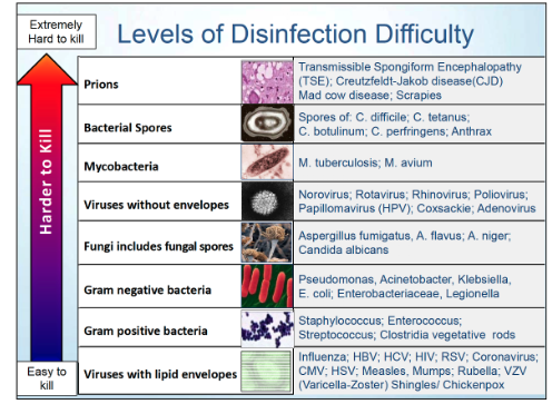
Prions are hardy pathogenic agents that remain at the top of Spalding’s Pathogen Hierarchy, as shown in Figure 1, for their heavy resistance to traditional disinfection and sterilization methods.7 They have been found to be readily transmissible and infectious in soil for at least two years.8 It is believed their hardiness comes from the significant folding of the proteins and potentially the amyloid fibers they generate.9 The absence of nucleic acids may also be a contributing factor to their overall resistance to traditional sterilization and decontamination methods.10 Traditional sterilization methods have been shown to be ineffective against prions (Table 2). Combination treatments of prions are required because no sterilization methods have resulted in 100% efficacy against these pathogens to date.11 To further highlight their resistance, a prion infected brain placed in formaldehyde for several decades was found to still provide infectious prions for placement of slides and for research purposes.
Figure 1: Levels of Disinfection Difficulty7
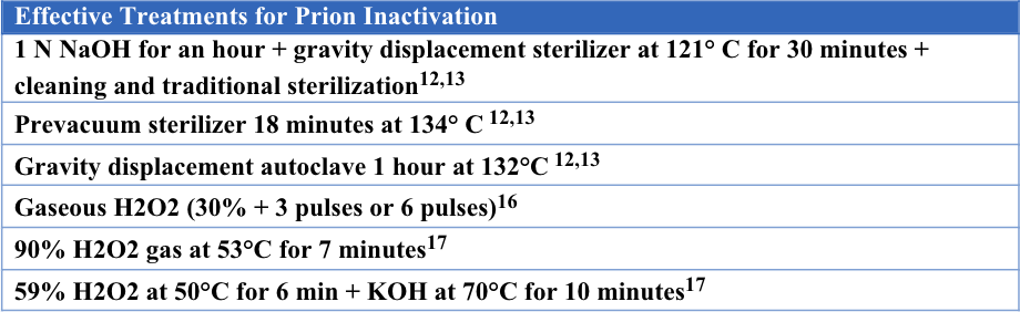
Current guidelines suggest that prion inactivation becomes significantly more challenging once the prions dry down and bind tightly to surfaces. For this reason, they should be kept moist through immersion in a liquid, such as water or a prionicidal detergent.12,13 One suggested method includes immersing the contaminated item in 1 N NaOH for an hour and then placing it into a gravity displacement sterilizer at 121° C for 30 minutes. After that, it should be cleaned again and undergo traditional sterilization. 12,13 Other options include using a prevacuum sterilizer autoclave for 18 minutes at 134° C or using a gravity displacement autoclave for 1 hour at 132°C. 12,13 Effective treatments for prion inactivation are described in Table 3. The methods that currently exist can be damaging to medical equipment or those employing the chemicals.
Table 2: Ineffective Sterilization Treatments for Prion Inactivation
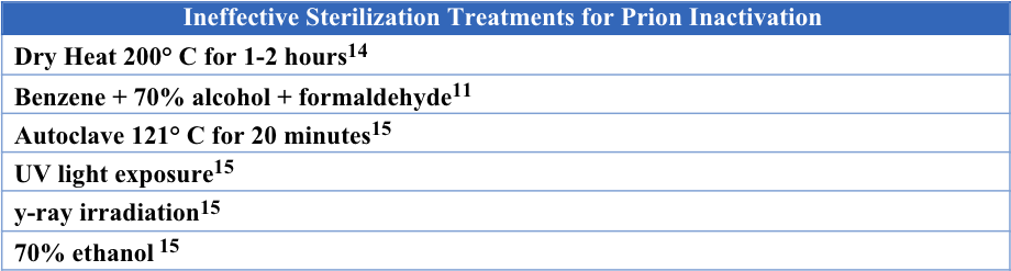
Table 3: Effective Treatments for Prion Inactivation
Newer studies have suggested the use of novel methods such as hydrogen peroxide vapor (HPV) for prion contaminated surgical instruments.16 Gaseous H2O2 is believed to be effective in prion disinfection by way of protein unfolding, protein fragmentation and a high sensitivity to proteolytic digestion.16 One study was conducted using the hamster adapted scrapie strain 263K, the BSE 6PB1 and BSE TGB1 strains in mice. Both 30% gaseous hydrogen peroxide with 3 and 6 pulses achieved greater than a 5.5 log reduction after testing on the 263K Scrapie Model and the 6PB1 BSE model (with 3 pulses only).16 A >5.3 log reduction was achieved with 30% gaseous hydrogen peroxide with 6 pulses on the TGB1 BSE model.16 Another study mirrored these results and found that VHP achieved a 4.5 log reduction on steel wires contaminated with hamster-adapted scrapie strain 263K.18 Liquid hydrogen peroxide fell short and was unsuccessful at disinfecting prion infected steel wires because it led to additional protein clumping.16 While high concentration hydrogen peroxide has undergone prion inactivation testing, further emphasis should be placed on testing lower concentration gaseous hydrogen peroxide for their potential to cause less degradation to equipment and to provide a safer alternative for staff.
Prion inactivation remains a challenge even with the best decontamination and sterilization technology available today. Risks to staff and corrosion to equipment remain prevalent concerns. The efficaciousness of products is still being debated about in the current literature as prions continue to win. Further research should be conducted on alternative methods and combination systems to provide the best chance at protecting patients from iatrogenic infections.
References
1. Prusiner SB. Prions. Proceedings of the National Academy of Sciences of the United States of America [Internet]. 1998 Nov 10;95(23):13363–83. Available from: https://www.ncbi.nlm.nih.gov/pubmed/9811807
2. Bae SE, Jung S, Kim HY, Son HS. Correlation analysis for the incubation period of prion disease. Prion [Internet]. 2012 Jul [cited 2019 Oct 27];6(3):276–81. Available from: https://www.ncbi.nlm.nih.gov/pmc/articles/PMC3399543/
3. Prions [Internet]. learn.genetics.utah.edu. University of Utah; Available from: https://learn.genetics.utah.edu/content/basics/prions
4. Beef cow with atypical bovine spongiform encephalopathy found in South Carolina [Internet]. American Veterinary Medical Association. 2023 [cited 2023 Oct 11]. Available from: https://www.avma.org/news/beef-cow-atypical-bovine-spongiform-encephalopathy-found-south-carolina#:~:text=Beef%20cow%20with%20atypical%20bovine%20spongiform%20encephalopathy%20found%20in%20South%20Carolina
5. Prion Diseases [Internet]. Centers for Disease Control and Prevention. Centers for Disease Control and Prevention; 2021. Available from: https://www.cdc.gov/prions/index.html
6. Bovine Spongiform Encephalopathy (BSE) Questions and Answers [Internet]. Center for Biologics Evaluation and Research. U.S. Food and Drug Administration; 2018 [cited 2023 Oct 11]. Available from: https://www.fda.gov/vaccines-blood-biologics/safety-availability-biologics/bovine-spongiform-encephalopathy-bse-questions-and-answers
7. Spaulding EH. Chemical disinfection of medical and surgical materials. In: Lawrence C, Block SS, editors. Disinfection, sterilization, and preservation. Philadelphia (PA): Lea & Febiger; 1968. p. 517–531
8. What are Prions? [Internet]. dwr.virginia.gov. Available from: https://dwr.virginia.gov/wildlife/diseases/cwd/what-are-prions/#:~:text=Most%20CWD%20research%20suggests%20incubation
9. Frazer J. Prions Are Forever [Internet]. Scientific American Blog Network. 2018. Available from: https://blogs.scientificamerican.com/artful-amoeba/prions-are-forever/
10. Sakudo A, Ano Y, Onodera T, Nitta K, Shintani H, Ikuta K, et al. Fundamentals of prions and their inactivation (Review). International Journal of Molecular Medicine. 2011 Jan 25;27(4):483–9.
11. McDonnell G, Burke P. The Challenge of Prion Decontamination. Clinical Infectious Diseases [Internet]. 2003 May 1 [cited 2020 Jun 23];36(9):1152–4. Available from: https://academic.oup.com/cid/article/36/9/1152/312297
12. Trapotsis A. The Challenge of Prion Sterilization [Internet]. Consolidated Sterilizer Systems. Available from: https://consteril.com/prion-sterilization-guide/
13. Rutala WA, Weber DJ. Guideline for Disinfection and Sterilization of Prion-Contaminated Medical Instruments. Infection Control & Hospital Epidemiology [Internet]. 2015 Jan 2;31(2):107–17. Available from: https://www.cdc.gov/infectioncontrol/pdf/guidelines/disinfection-guidelines.pdf
14. Jung, M. J., Pistolesi, D., & Panà, A. (2003). Prions, prion diseases and decontamination. Igiene e sanita pubblica, 59(5), 331–344.
15. Sakudo A. Inactivation Methods for Prions. Current Issues in Molecular Biology. 2020;36:23–32.
16. Fichet G, Antloga K, Comoy E, Deslys JP, McDonnell G. Prion inactivation using a new gaseous hydrogen peroxide sterilisation process. Journal of Hospital Infection. 2007;67(3):278–86.
17. Sakudo A, Yamashiro R, Onodera T. Recent Advances in Prion Inactivation by Plasma Sterilizer. International Journal of Molecular Sciences [Internet]. 2022 Sep 6 [cited 2023 Oct 11];23(18):10241. Available from: https://www.mdpi.com/1422-0067/23/18/10241
18. Fichet G, Comoy E, Duval C, Antloga K, Dehen C, Charbonnier A, et al. Novel methods for disinfection of prion-contaminated medical devices. The Lancet. 2004 Aug 7;364(9433):521–6.